EYEPLUS EYE CLINIC GLAUCOMA EYEPLUS EYE CLINIC GLAUCOMA EYEPLUS EYE CLINIC GLAUCOMA EYEPLUS EYE CLINIC GLAUCOMA EYEPLUS EYE CLINIC GLAUCOMA

Glaucoma Guide:
Symptoms, Diagnosis, and Treatment

glaucoma_hero_img

Glaucoma is a serious eye condition that can gradually damage the optic nerve and lead to permanent vision
loss if left untreated. One of the most concerning aspects of glaucoma is that it often progresses without noticeable
symptoms in the early stages, making early detection essential.

This comprehensive guide is designed to help you understand glaucoma symptoms, causes, and risk factors, as well as how the
condition is identified through accurate glaucoma diagnosis. It also explains the latest glaucoma treatment options
available, including both medical and surgical approaches.

At EYE Plus Eye Clinic, we focus on early diagnosis and personalized care to help protect your vision. Whether
you are experiencing symptoms or simply want to learn more, this guide will provide clear and reliable information for
international patients seeking safe and effective eye care in Korea.

Why Is It Called the “Silent Thief of Sight”?

What Is Glaucoma?

Glaucoma is one of the leading causes of blindness worldwide. It is a progressive eye disease in which the optic nerve is gradually damaged, causing the field of vision to slowly narrow over time.

In many cases, glaucoma develops without noticeable pain or early symptoms. Because patients often do not realize the condition until vision loss has already progressed, it is commonly referred to as the “silent thief of sight”.

A common misconception is that glaucoma only occurs when eye pressure is high. However, normal eye pressure does not guarantee safety. Glaucoma cannot be ruled out based on intraocular pressure alone, and a more comprehensive evaluation is necessary for accurate diagnosis.

Why You Should Not Feel Safe Even with Normal Eye Pressure (Normal Tension Glaucoma)

안압상승 사진

Normal tension glaucoma (NTG) is a type of glaucoma where optic nerve damage occurs even though intraocular pressure remains within the normal range (typically around 10–21 mmHg). This condition is especially common among Asian patients.

Even within the “normal” range, susceptibility to glaucoma varies from person to person. Some individuals may develop glaucoma at relatively low eye pressure, while others can tolerate higher pressure without damage. This means that eye pressure alone is not enough to assess glaucoma risk.

The main cause of normal tension glaucoma is believed to be related to structural vulnerability of the optic nerve and impaired blood flow, rather than pressure itself.

Therefore, accurate diagnosis requires a comprehensive approach, including:

  • Optic nerve evaluation

  • Difficulty focusing due to decreased vision

  • OCT (Optical Coherence Tomography) imaging

  • Visual field testing

These tests help determine the presence of optic nerve damage and the progression of glaucoma more precisely.

Possible symptoms may include:

  • Eye discomfort or pressure sensation

  • Difficulty focusing due to decreased vision

  • Blurred vision

  • Headaches

  • Nausea or vomiting (in advanced cases)

Because glaucoma can progress silently, regular eye examinations are essential, even if you do not experience clear symptoms or have normal eye pressure.

Advanced Glaucoma Diagnosis at EYE Plus Eye Clinic

Glaucoma is a condition where early and accurate diagnosis is critical, as damage to the optic nerve is irreversible once it occurs. The earlier glaucoma is detected, the better the chances of preserving vision.

At EYE Plus Eye Clinic, we go beyond simple eye pressure measurements. Using advanced diagnostic equipment and a comprehensive evaluation system, we analyze both the structure and function of the optic nerve in detail.

This allows us to detect glaucoma even in its earliest stages, including cases without obvious symptoms, and to accurately assess the risk of progression.

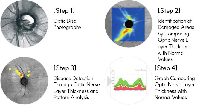
Cirrus HD-OCT for Detailed Optic Nerve Analysis

Unlike basic examinations that only assess the overall shape of the optic nerve, Cirrus HD-OCT provides a microscopic analysis of the retinal nerve fiber layer (RNFL) and ganglion cell layer.

This advanced OCT glaucoma test can detect subtle structural changes before they appear in visual field tests, making it highly effective for early diagnosis (pre-perimetric glaucoma).

It also allows for precise comparison with previous results, enabling continuous monitoring of disease progression rather than a one-time diagnosis.

녹내장 정밀 검사 단계 이미지 — 시신경 유두촬영, 시신경층 두께 및 패턴 분석, 정상인과 비교한 손상 부위 표시, 그래프 분석을 통해 시신경 손상 여부를 진단하는 과정을 설명함.

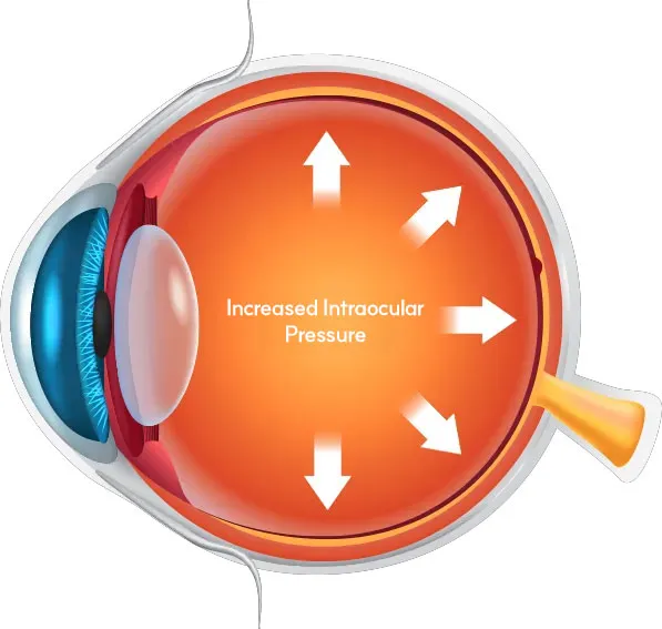
Intraocular Pressure Test (Basic Screening Test)

The intraocular pressure (IOP) test is the most basic glaucoma screening method. Normal eye pressure typically ranges from 10 to 21 mmHg, which helps maintain the shape of the eye.

However, normal eye pressure does not always mean the eye is healthy. If elevated pressure or suspicious findings are detected, additional tests such as OCT or visual field testing are required for accurate diagnosis.

비접촉 안압 측정기(Nidek NT-510) 장비 이미지

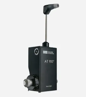
Goldmann Applanation Tonometry (Gold Standard Eye Pressure Test)

While basic air-puff tests are useful for screening, they may vary depending on corneal thickness and eye condition.

Goldmann applanation tonometry is considered the most accurate method for measuring eye pressure and is widely used as the gold standard in major hospitals.

This test involves gently contacting the cornea with a specialized prism, allowing for highly precise measurement of true intraocular pressure, especially when a detailed diagnosis is needed.

골드만 압평안압계 장비 이미지

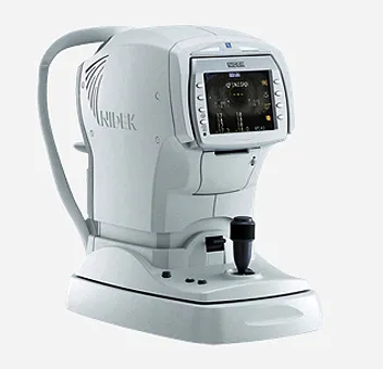
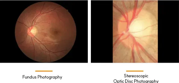
Non-Dilated Fundus Photography (Optic Nerve Examination)

Non-dilated fundus imaging allows detailed observation of the optic nerve head without discomfort, enabling early detection of structural changes.

Visual Field Test

Glaucoma primarily affects the optic nerve head, making its evaluation essential.

With non-dilated fundus photography, the optic nerve can be clearly examined without the need for pupil dilation, reducing patient discomfort.

This test helps identify:

  • Changes in the optic nerve structure

  • Abnormal cupping or boundaries

  • Early signs of glaucoma-related damage

It enables fast, accurate detection and monitoring of structural changes associated with glaucoma.

녹내장 검사 안저사진과 시신경유두 입체촬영 이미지 — 시신경 손상 여부와 시야 결손 진행 정도를 확인하기 위한 정밀 안과 검사 과정.

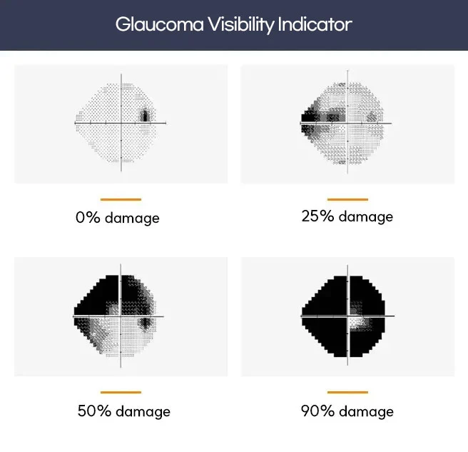
Visual Field Test (Checking the Progression of Glaucoma)

The visual field test evaluates how much your peripheral (side) vision has been affected by glaucoma.

As the optic nerve becomes damaged, the range of vision gradually narrows. This test measures those changes in a quantitative and visual format, allowing doctors to:

  • Detect blind spots (scotomas)

  • Assess the severity of glaucoma

  • Monitor disease progression over time

By comparing results over time, it plays a crucial role in treatment planning and long-term management of glaucoma.

녹내장 시야 지표 비교 이미지 — 시신경 손상 정도에 따른 시야 손실 변화를 0%, 20%, 50%, 90% 단계로 나타내어 녹내장의 진행 상태를 시각적으로 보여줌.

Stage-Based Customized Glaucoma Treatment Solutions

The key to effective glaucoma treatment is not relying on a single method, but taking a step-by-step, personalized approach based on each patient’s condition.

Because glaucoma progression, causes, and the degree of optic nerve damage vary from person to person, treatment should follow a structured plan—from medication → laser treatment → surgical intervention—depending on disease severity and response.

  • 안약 점안 아이콘

    Medication Treatment (Eye Drops)

    Eye drops are the first-line treatment for glaucoma. These medications help lower intraocular pressure (IOP) and slow the progression of optic nerve damage.

    Regular and consistent use is essential, as glaucoma requires long-term management rather than short-term relief. In some cases, medication alone may not be sufficient, and treatment may need to be adjusted based on follow-up examinations and pressure control.

  • 레이저 치료 아이콘

    Laser Treatment for Glaucoma

    When eye drops are not enough to control intraocular pressure, laser treatment (such as SLT) may be recommended.

    Laser procedures work by improving the drainage of aqueous humor (fluid) inside the eye, helping to lower eye pressure more effectively.
    They are generally minimally invasive, do not require incisions, and involve relatively quick recovery times.

    However, the effectiveness and duration of results may vary depending on the individual, so careful evaluation is necessary.

  • 섬유주절제술 아이콘

    Trabeculectomy

    If both medication and laser treatment fail to adequately control intraocular pressure, trabeculectomy may be considered.

    This surgical procedure creates a new drainage pathway (bleb) that allows fluid to exit the eye, thereby reducing intraocular pressure more effectively.

    Trabeculectomy is typically recommended for advanced or progressive glaucoma, where stronger and more sustained pressure control is required to prevent further vision loss.

Dry Eye Syndrome FAQ

Consultation Request

Please leave your questions for EYEPLUS,
and our staff will contact you after reviewing them.