EYEPLUS EYE CLINIC RETINAL DISEASE EYEPLUS EYE CLINIC RETINAL DISEASE EYEPLUS EYE CLINIC RETINAL DISEASE

Retinal Disease Types and Treatment

retinal disease

Retinal diseases are conditions that affect the retina, the light-sensitive tissue at the back
of the eye that plays a critical role in vision. When the retina is damaged,
the eye cannot properly process visual information, which can lead to blurred vision, visual distortion, or even vision loss.

Common types of retinal disease include floaters, macular degeneration, retinal detachment, retinal tears,
and diabetic retinopathy. These retinal disorders can develop gradually or suddenly,
and symptoms may be subtle in the early stages.

Because retinal tissue has very limited ability to recover once damaged, early diagnosis and timely treatment
are essential. Detecting retinal diseases at an early stage can significantly
improve treatment outcomes and help prevent permanent vision loss.

What Is the Retina?

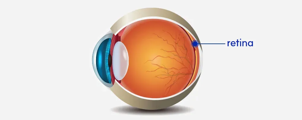
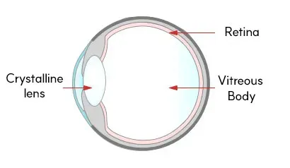
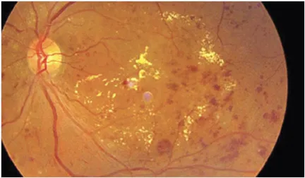
The retina is a thin layer of nerve tissue located at the back of the eye, playing a crucial role in vision. It functions like a camera sensor, receiving light from outside the eye, converting it into visual information, and transmitting it to the brain through the optic nerve.

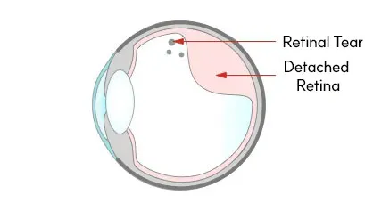
When the retina is damaged, it can lead not only to blurred vision but also to distortion or even permanent vision loss. This is why early detection and proper management of retinal conditions are essential.

Light Detection and Signal Conversion

The retina’s primary function is to detect incoming light and convert it into electrical signals. These signals are transmitted through the optic nerve to the brain, where they are interpreted as visual images.

If this process is disrupted, vision may become blurred or distorted, affecting overall visual clarity.

Color and Shape Recognition

The retina contains specialized cells called photoreceptors, including cone cells and rod cells.

  • Cone cells are responsible for color vision and fine detail

  • Rod cells detect brightness and help recognize shapes, especially in low light

Together, these cells allow us to distinguish colors, brightness, and object outlines clearly. Damage to these cells may result in reduced color perception or blurred vision.

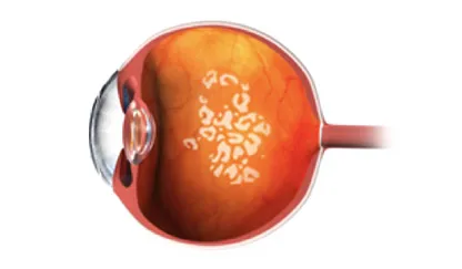
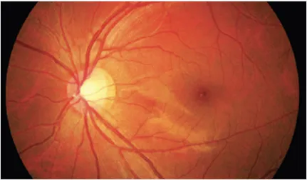
High-Resolution Vision at the Macula

At the center of the retina lies the macula, a small but critical area responsible for sharp and detailed central vision. Although it occupies only a small portion of the retina, it accounts for the majority of high-quality vision.

The macula is essential for activities such as reading, recognizing faces, and performing precise tasks. Damage to this area can significantly impact daily life by reducing visual clarity and detail perception.

Floaters (Vitreous Floaters)

Eye floaters (vitreous floaters) are visual disturbances that appear as small dots, lines, or shadow-like shapes drifting across your field of vision. These occur when the vitreous gel inside the eye changes and casts shadows on the retina.

In most cases, floaters are a natural part of aging. However, in some situations, they may indicate underlying retinal problems, so understanding the difference is important.

Normal Vitreous
정상 유리체 이미지
정상 시야 이미지 — 맑은 하늘과 구름이 깨끗하게 보이는 건강한 눈의 시야 상태를 표현함.

Normal Vision

Vitreous with Floaters
비문증 유리체 이미지
비문증 시야 이미지 — 하늘을 볼 때 떠다니는 점이나 실 같은 검은 그림자가 보이는 비문증 증상을 시각적으로 표현함.

Vitreous Floaters

Physiological Floaters (Age-Related Floaters)

Normal floaters are commonly caused by age-related changes in the vitreous. As we get older, the vitreous becomes more liquefied and can form tiny clumps, which appear as floaters.

  • 체크 아이콘

    Develop gradually over time

  • 체크 아이콘

    Little to no change in size or number

  • 체크 아이콘

    Not associated with vision loss or eye pain

In most cases, these floaters do not require treatment. The brain often adapts over time, making them less noticeable.

Pathological Floaters That Require Examination

Certain symptoms may indicate a more serious condition and require immediate eye examination:

  • 체크 아이콘

    Sudden increase in floaters

  • 체크 아이콘

    Flashes of light (photopsia)

  • 체크 아이콘

    A shadow or “curtain” effect in part of the vision

These signs may be associated with retinal tear, retinal detachment, or vitreous hemorrhage.

If floaters and flashes occur together, it is especially important not to assume it is simple aging. A comprehensive retinal examination is necessary to determine the cause and prevent potential vision loss.

Retinal Detachment and Retinal Tear

Retinal detachment and retinal tear are serious eye conditions that require urgent attention. These two conditions are closely related—retinal tears can progress to retinal detachment if left untreated.

If detected early at the tear stage, progression can often be prevented. However, once detachment occurs, the risk of permanent vision loss increases significantly. Prompt diagnosis and treatment are essential.

Retinal Detachment

Retinal detachment occurs when the retina separates from the back of the eye, disrupting its normal function.

  • A curtain-like shadow over part of the vision

  • Sudden decrease in vision

  • Dark shadows or loss of peripheral vision

This condition blocks the retina’s blood supply, and the longer it is left untreated, the lower the chance of visual recovery.

Surgical treatment is usually required, often using advanced techniques (such as vitrectomy or specialized surgical systems). Early intervention is critical to preserving vision.

Retinal Tear

A retinal tear is a small break or hole in the retina and is often the early stage before detachment.

  • May occur before the retina fully detaches

  • Often associated with sudden floaters or flashes of light

At this stage, laser treatment (laser photocoagulation) can seal the tear and prevent progression to retinal detachment.

If symptoms such as a sudden increase in floaters or flashes appear, immediate examination is necessary. Early diagnosis and prompt laser treatment play a key role in protecting vision.

Normal Retina
정상 안구 단면도 이미지
Retinal Tear
망막열공 단면도 이미지 — 망막에 구멍이 생기거나 일부가 박리되어 시야 손상과 시력 저하를 유발하는 망막열공의 진행 상태를 보여줌.

Macular Degeneration

Macular Degeneration is a condition that affects the macula, the central part of the retina responsible for sharp, detailed vision. It primarily causes central vision loss and can significantly impact daily activities such as reading and recognizing faces.

In the early stages, it may be mistaken for presbyopia or eye fatigue, but as it progresses, it can lead to noticeable visual impairment. Because the macula plays a key role in vision, early detection and precise diagnosis are essential.

Main Symptoms of Macular Degeneration

  • 체크 아이콘

    Straight lines appear bent or distorted

  • 체크 아이콘

    Dark or blurred spot in the center of vision (central vision loss)

  • 체크 아이콘

    Overall blurry or hazy vision

  • 체크 아이콘

    Gradual decline in visual clarity, even with glasses

What Is the Amsler Grid Test?

The Amsler grid test is a simple and effective way to check the condition of the macula. It helps detect early signs of distorted vision or central vision problems.

This test can be easily performed at home and is useful for identifying subtle changes in vision before symptoms become severe.

암슬러 격자 이미지

[How to Perform the Amsler Grid Test]

1. In a well-lit area, wear your glasses or reading glasses and hold the grid at eye level.

2. Cover one eye and focus on the center dot with the other eye.

3. Check whether the grid lines appear bent, distorted, blurred, or missing.

4. Repeat the same steps with the other eye.

Symptoms That Require Immediate Eye Examination

If you notice any of the following symptoms, visit an eye clinic as soon as possible:

Straight lines appear wavy or distorted
Parts of your vision look missing or like a dark spot
Objects appear warped or irregular in shape
These may be warning signs of macular degeneration progression and require prompt evaluation to prevent further vision loss.

Diabetic Retinopathy

Diabetic retinopathy is one of the most common complications of diabetes and a leading cause of vision loss. When high blood sugar levels persist, they damage the small blood vessels in the retina, leading to reduced vision over time.

As the condition progresses, weakened blood vessels may leak or bleed, causing swelling and further retinal damage. In severe cases, it can result in significant vision loss or even blindness.

A critical concern is that early stages often have no noticeable symptoms. Even if vision seems normal, retinal damage may already be developing silently.

This is why regular eye examinations are essential. Early detection through routine check-ups allows timely treatment and helps slow disease progression. Along with proper blood sugar control, consistent eye care plays a key role in protecting vision.

Normal Fundus
정상 안저 이미지 — 시신경과 망막 혈관이 뚜렷하게 보이는 건강한 눈의 안저 사진.
Diabetic Retinopathy Fundus
당뇨망막병증 안저 이미지 — 망막 내 출혈과 삼출물이 관찰되는 당뇨 합병증으로, 시력 저하를 유발하는 당뇨망막병증의 특징적인 소견을 보여줌.

Advanced Retinal Examination System

The outcome of retinal disease often depends on how early it is detected. At EYE Plus Eye Clinic, we use an advanced retinal examination system to identify both structural and functional retinal abnormalities at an early stage.

Rather than relying on a basic vision test alone, we evaluate the entire retina—including the central and peripheral areas—to support accurate diagnosis and timely treatment.

정밀 검사기 사진

Wide-Field Fundus Camera (Optos)

The wide-field fundus camera (Optos) can capture up to approximately 200 degrees of the retina in a short time, even without pupil dilation.

This makes it especially useful for detecting:

  • 체크 아이콘

    Peripheral retinal abnormalities

  • 체크 아이콘

    Early retinal tears or retinal detachment

  • 체크 아이콘

    Small vascular changes that may otherwise be missed

  • Because the examination is fast and causes less discomfort, it also helps reduce patient burden during retinal screening.

Optical Coherence Tomography (OCT)

OCT (Optical Coherence Tomography) scans the retina in cross-section with high precision, allowing doctors to detect subtle abnormalities that may not be visible during a standard eye exam.

A retinal OCT scan is particularly useful for evaluating:

  • 체크 아이콘

    Macular degeneration

  • 체크 아이콘

    Diabetic retinopathy

  • 체크 아이콘

    Macular edema and retinal thickness changes

  • This test provides objective, detailed information about retinal structure, helping determine whether treatment is needed and how the disease is progressing.

Who Should Get a Detailed Retinal Examination?

A detailed retinal examination is recommended for individuals at higher risk of retinal disease or those experiencing changes in vision. Regular retina checkups are essential for early detection, as many retinal conditions develop without obvious symptoms in the early stages.

You should consider a retinal exam if you fall into any of the following groups:

  • 체크 아이콘

    People with diabetes or high blood pressure

    These conditions can damage the small blood vessels in the retina, even without noticeable symptoms. Regular monitoring is important to prevent complications.
  • 체크 아이콘

    People experiencing vision changes

    Symptoms such as narrowing vision, blurred areas, or a “blocked” feeling in part of your vision may be early signs of retinal disease.
  • 체크 아이콘

    People with decreased vision not improved by glasses

    If your vision does not improve after changing glasses, it may be related to retinal abnormalities rather than simple refractive error.
  • 체크 아이콘

    Adults over the age of 55

    The risk of retinal diseases such as macular degeneration increases with age, making regular eye examinations more important.
  • 체크 아이콘

    People with high myopia (severe nearsightedness)

    A longer eye length can make the retina thinner and more vulnerable to conditions such as retinal tears or detachment.
  • 체크 아이콘

    People who see distorted or warped images

    If straight lines appear bent or objects look distorted, it may indicate a problem in the macula or central retina.

* A more accurate diagnosis can be found through a specialized examination. Please feel free to proceed with the consultation or appointment through the button below.

FAQ About Retinal Disease

상담신청

아이플러스에
궁금하신 점을 남겨주시면
담당자 확인 후 연락드리겠습니다.