EYEPLUS IMPLANTABLE CONTACT LENS EYEPLUS IMPLANTABLE CONTACT LENS EYEPLUS IMPLANTABLE CONTACT LENS

Implantable Collamer Lens (ICL) Guide:
Types, Benefits, Risks, and Safety Facts

ICL surgery

Implantable Collamer Lens (ICL) is an advanced vision correction option for patients who may not be suitable candidates for laser procedures
such as LASIK or LASEK. Unlike laser surgery, ICL corrects vision by placing a biocompatible lens inside the eye without removing corneal tissue.

This page is designed as a complete guide to implantable collamer lens (ICL) surgery, covering the questions patients most often ask before treatment.
You will find clear information about lens types, safety, recovery process, cost, and possible side effects.

At EYE Plus Eye Clinic, ICL treatment is planned based on detailed eye examinations and extensive clinical experience in refractive surgery.
This guide reflects the clinic’s practical expertise and is intended to help patients better understand whether implantable collamer lens surgery is the right option for their eyes.

Whether you are considering an alternative to laser vision correction or exploring options for high myopia and thin corneas,
this guide will help you understand how ICL works and what to expect beforemaking a decision.

When Is It Chosen Instead of LASIK or LASEK?

What Is Implantable Collamer Lens (ICL) Surgery?

Implantable Collamer Lens (ICL) surgery corrects refractive errors by placing a biocompatible lens inside the eye, typically behind the iris and in front of the natural lens, without cutting or reshaping the cornea. Unlike LASIK or LASEK, the corneal surface remains untouched.

Because the cornea is preserved, implantable collamer lens surgery is often recommended for patients with thin corneas, very high myopia, severe astigmatism, or dry eyes that make laser vision correction less suitable.

ICL is also considered by patients who wish to avoid permanent corneal tissue removal. Since the implanted lens can be removed or replaced if necessary, it offers a reversible alternative to laser procedures.

When comparing ICL vs LASIK or ICL vs LASEK, the most suitable choice depends on corneal condition, refractive error, and internal eye structure.

Key Comparison of Vision Correction Procedures: LASIK vs LASEK vs ICL

Category LASIK LASEK ICL
Procedure
Method
Corneal flap creation (approx. 20 mm incision) Removal of corneal epithelium Cornea preserved, lens implanted
Pain /
Discomfort
Relatively mild Possible discomfort for 2–3 days in early recovery Generally mild, varies by patient
Recovery
Speed
Daily activities possible the next day Protective lens for about 3 days, vision stabilizes in 1–2 weeks Immediate recovery in many cases
Resistance to
External Impact
Flap displacement risk exists Strong after epithelial healing No corneal damage
Dry Eye Risk Possible due to corneal nerve disruption Relatively lower Does not usually induce dry eye
Recommended
For
Patients with suitable corneal thickness seeking fast recovery Patients with thin corneas or active lifestyles Patients with very high myopia, astigmatism, or thin corneas

The most suitable vision correction method depends on corneal thickness, degree of myopia, anterior chamber depth, and individual eye structure.

👉 Curious about iPlus Eye Clinic’s proven all-laser LASIK procedure? [Learn more about LASIK]

👉 Interested in iPlus Eye Clinic’s LASEK with the FROST COOLING system for minimized discomfort? [Learn more about LASEK]

Who Is a Good Candidate for Implantable Collamer Lens (ICL) Surgery?

Implantable Collamer Lens (ICL) surgery is often recommended for patients who are not ideal candidates for laser vision correction. Because the cornea is preserved, ICL can be a safer option when LASIK or LASEK is limited by corneal thickness, refractive range, or eye condition.

ICL may be especially suitable for patients with high myopia above approximately -6.00 diopters, thin corneas that make laser surgery unsafe, or severe dry eye that raises concerns about corneal laser procedures. It is also considered for patients who prefer not to alter corneal tissue permanently.

Patients who have experienced regression after previous vision correction surgery may also be candidates for ICL as an alternative corrective option.

Recommended for Patients Who:

  • 체크 아이콘

    Have thin corneas and cannot safely undergo LASIK or LASEK
  • 체크 아이콘

    Have very high myopia that exceeds the safe correction range of laser surgery
  • 체크 아이콘

    Prefer a reversible procedure that preserves natural corneal tissue
  • 체크 아이콘

    Are concerned about dry eye, glare, or night vision issues after laser correction
  • 체크 아이콘

    Have irregular corneal shape or corneal abnormalities
  • 체크 아이콘

    Need an alternative after previous laser vision correction regression

Lens Types Explained: Finding the Right Implantable Collamer Lens for Your Eyes

ICL lenses are broadly divided by placement position into anterior chamber lenses and posterior chamber lenses. The choice depends on where the lens is implanted inside the eye and how it is stabilized.

Anterior chamber lenses are positioned in front of the iris, while posterior chamber lenses are placed behind the iris and in front of the natural lens. Modern ICL surgery most commonly uses posterior chamber lenses because of their improved long-term safety profile.

체크 아이콘

Front lens

Insert the lens in front of the iris The lens is fixed directly to the iris, so the fixing power is good

안내렌즈삽입술 전방렌즈 삽입 예시이미지

알티산 렌즈 이미지

Artisan

First U.S. FDA Approved a wide range of vision correction Minimize glare and light smudging

알티플렉스 렌즈 이미지

Artiplus

a quick recovery Minimize dry eyes Reducing cataract side effects
Rear lens

Insert the lens behind the iris, no iris incision required and high stability

안내렌즈삽입술 후방렌즈 삽입 예시이미지

EVO+ AQUA ICL 렌즈 이미지

EVO+ AQUA ICL

excellent corneal preservation
high safety a quick recovery

Anterior Chamber Lens Types and Characteristics

Anterior chamber lenses are placed in front of the iris and secured within the anterior chamber of the eye. Common examples include iris-fixated lenses such as Artisan and Artiflex.

Artisan was one of the earliest widely used implantable lens types and provides a broad range of vision correction. Artiflex was later developed as a flexible version designed to allow faster recovery and improved comfort after surgery.

Because anterior chamber lenses are positioned closer to the cornea, they may increase the long-term risk of corneal endothelial cell loss in some patients. To reduce this risk and improve long-term safety, posterior chamber lenses are now more commonly used in modern ICL surgery.

Posterior Chamber Lens Types and Recommended Options

Posterior chamber lenses are implanted behind the iris, in front of the natural crystalline lens. This position avoids corneal cutting and reduces direct impact on corneal endothelial cells.

Modern posterior chamber ICLs, such as EVO+ AQUA ICL, are now the most widely recommended option because they offer strong corneal preservation, high safety, and fast recovery.

For mild to moderate astigmatism, EVO+ AQUA ICL is commonly used. This lens is made from biocompatible Collamer material, which is soft, flexible, and highly compatible with the eye.

For high astigmatism, Toric ICL is recommended. Toric lenses correct both severe myopia and astigmatism simultaneously, providing sharper and more stable visual outcomes.

Accurate lens selection is essential in ICL surgery. The most appropriate lens type is determined through detailed preoperative measurements, including refractive error, anterior chamber depth, and eye anatomy.

Potential Side Effects of ICL Surgery and Medical Facts

When considering Implantable Collamer Lens (ICL) surgery, patients often worry about possible complications. The most common concerns involve endothelial cell loss, cataract risk, and increased eye pressure. These risks are important to understand, but modern ICL technology and proper screening have significantly improved long-term safety.

At EYE Plus Eye Clinic, every ICL procedure is planned through detailed diagnostic testing to minimize risks before surgery. Accurate measurements and careful patient selection are essential for safe and stable outcomes.

Endothelial Cell Loss: “Can I Lose Corneal Cells After ICL Surgery?

Reference One of the most common concerns about implantable collamer lens side effects is whether endothelial cells continue decreasing after surgery and may eventually damage vision.

Medical evidence shows reassuring results. According to long-term published clinical research, including a ' 10-year study on ICL safety', endothelial cell reduction after properly performed ICL surgery is not significantly different from the natural decrease that occurs with aging.

This means that when patients are carefully screened and appropriate surgical standards are followed, abnormal rapid endothelial cell loss is uncommon.

At EYE Plus Eye Clinic, endothelial cell density is measured both before and after surgery using specular microscopy (SP examination), and long-term follow-up is used to monitor corneal safety. If the internal eye space is insufficient or safety criteria are not met, surgery is not recommended.

Sources: Ten-Year Clinical Outcomes of V4c Implantable Collamer Lens Implantation (American Journal of Ophthalmology, 2024)

Cataract Risk: “What If the Lens Touches My Natural Lens?”

Some patients worry that the implanted lens may touch the natural crystalline lens and cause cataracts.

This complication is mainly related to incorrect lens sizing or insufficient space inside the eye. If the implanted lens sits too close to the natural lens, continuous contact or friction may increase cataract risk over time.

This is why vault is critical in ICL surgery. Vault refers to the safe distance maintained between the implanted lens and the natural lens.

At EYE Plus Eye Clinic, precise lens sizing is determined in 0.01 mm increments using advanced diagnostic measurements, including UBM (ultrasound biomicroscopy), which directly measures internal eye dimensions. This allows surgeons to select lens size based on actual anatomical space rather than visible external estimates.

By maintaining a proper vault, the risk of cataract formation can be reduced to nearly zero in well-selected patients.

Increased Eye Pressure and Glaucoma: “Can ICL Surgery Raise Eye Pressure?”

Another common concern is whether ICL surgery may block fluid circulation inside the eye and cause increased eye pressure or glaucoma.

In earlier lens designs, impaired aqueous fluid flow could occasionally contribute to pressure elevation. Some older lens models required additional procedures to create fluid pathways.

Modern EVO ICL lenses have solved this issue with Centra Flow technology. This design includes a central flow channel in the lens that allows natural aqueous circulation without additional surgical openings.

Because of this innovation, the risk of sudden eye pressure rise after surgery has been significantly
Before surgery, EYE Plus Eye Clinic carefully evaluates intraocular pressure, anterior chamber depth, and internal eye anatomy. If there is any elevated glaucoma risk, surgery is not performed.

With modern EVO ICL technology and strict screening standards, concerns related to glaucoma and pressure-related complications are greatly minimized.

Advanced Pre-Surgery Examination Process for Implantable Collamer Lens (ICL) Surgery

The success of Implantable Collamer Lens (ICL) surgery is determined before the operation begins. Even very small measurement errors can affect long-term safety and visual outcomes, which is why EYE Plus Eye Clinic goes beyond standard vision testing to analyze the internal structure of the eye in detail.

For safe ICL surgery, precise lens sizing is essential. This requires not only measuring refractive error, but also evaluating vault prediction, anterior chamber depth, internal eye space, and anatomical compatibility before selecting the lens.

ARK: Auto Refractor & Keratometer

an examination that measures the refractive power of the eye, such as myopia/rawness/astigmatism, and corneal curvature

IOP: Intraocular Pressure

An important test for early detection of eye diseases such as glaucoma by measuring eye pressure (waterproof pressure)

Pentacam HR: 3D Anterior Eye Examination

The front part of the cornea and eyeball is precisely photographed in 3D to analyze the shape, thickness, and depth of the cornea

Galilei G4: Comprehensive
Cornea & Anterior Eye Exam

Comprehensive cornea & anterior eye exam analyzing corneal topography, thickness, front and back surface shape, and anterior chamber depth

OCT: Optical coherence tomography

High-resolution retinal imaging scan for precise diagnosis of retinal, optic nerve, and corneal conditions using light-based tomography

OPTOS: Wide-Field Retinal Imaging

Advanced wide-field retinal imaging that captures up to 80% of the retina without pupil dilation for early detection of retinal diseases

Vault: The Safe Distance Between the Implanted Lens and the Natural Lens

In ICL surgery, vault refers to the microscopic space between the implanted lens and the natural crystalline lens inside the eye. This distance is one of the most important safety indicators after surgery.

Proper vault is essential because the implanted lens must remain at a stable distance from the natural lens. If the gap is too narrow, the implanted lens may touch the natural lens and increase cataract risk. If the gap is too wide, it may interfere with aqueous fluid circulation and raise intraocular pressure.

The generally accepted ideal vault range is approximately 250–750 μm, which allows both safe lens positioning and natural fluid flow.

Vault conditions are commonly interpreted as follows

  • 체크 아이콘

    Ideal Vault (250–750 μm)
    Safest range for long-term stability and normal fluid circulation

  • 체크 아이콘

    Low Vault (below 250 μm)
    Increased risk of lens contact with the natural lens, which may contribute to cataract formation

  • 체크 아이콘

    High Vault (above 1000 μm)
    May compress surrounding eye structures, disrupt fluid drainage, and increase eye pressure or glaucoma risk

Beyond What Is Visible: Limitations of WTW and the Accuracy of UBM Measurement

One of the most important parts of ICL lens sizing is selecting a lens that precisely fits the actual internal eye space. Many clinics use WTW (white-to-white) measurement, which measures the visible horizontal corneal diameter from the outside.

Although WTW is simple and convenient, it does not fully reflect the true internal anatomy of the eye. External eye width does not always match the internal space where the implanted lens will actually sit.

In reality, the most important measurement is the internal sulcus-to-sulcus (STS) distance — the actual space behind the iris where the lens is positioned. This cannot be accurately determined through external observation alone.

Reference

Clinical studies have reported that
WTW-based lens size prediction accuracy is limited to approximately 83%meaning sizing error remains possible in about 1–2 out of 10 cases. More precise methods such as UBM improve vault prediction and long-term ICL safety.

By combining UBM with comprehensive anatomical analysis, EYE Plus Eye Clinic improves lens selection precision and minimizes preventable sizing-related complications.

At EYE Plus Eye Clinic, UBM (Ultrasound Biomicroscopy) is used to directly measure internal eye structures, including STS distance. This allows much more accurate lens sizing and helps reduce complications caused by oversized or undersized lenses.

High-Precision Implantable Collamer Lens Surgery Procedure

For many first-time patients, Implantable Collamer Lens (ICL) surgery may sound complex because it involves placing a lens inside the eye. In reality, it is a highly precise but minimally invasive procedure designed to reduce discomfort while maintaining a high level of safety.

At EYE Plus Eye Clinic, implantable collamer lens surgery is typically completed within about 10–15 minutes under topical anesthesia. Because the cornea is not widely cut and only a micro incision is required, pain during surgery is minimal and recovery is generally comfortable.

Rather than focusing on speed alone, EYE Plus prioritizes accuracy and safety throughout every stage of the procedure so patients can feel reassured from beginning to end.

  • icl_step1

    Step 1. Topical Anesthesia and Micro Incision

    Implantable collamer lens surgery begins with topical anesthesia using anesthetic eye drops rather than injection anesthesia.

    Because no needle anesthesia is required, patients can begin surgery without injection-related discomfort. After anesthesia takes effect, a very small incision of approximately 2–3 mm is made to safely insert the lens into the eye.

    This minimally invasive approach helps reduce tissue disruption, lowers discomfort during surgery, and supports faster healing after the procedure.

  • icl_step2

    Step 2. Insertion of a Customized Implantable Collamer Lens

    The customized implantable collamer lens selected during the pre-surgery examination is carefully inserted into the safe space behind the iris.

    Each lens is individually chosen based on the patient’s eye structure, refractive error, and internal measurements. Because the lens is tailored to the patient’s anatomy, it can be positioned accurately while minimizing unnecessary irritation to the cornea and natural crystalline lens.

    This customized placement is one of the key factors that allows stable and precise long-term vision correction.

  • icl_step3

    Step 3. Lens Position Stabilization and Final Safety Check

    After the lens is inserted, the surgeon carefully confirms that it is correctly centered and safely positioned behind the iris.

    Any remaining surgical solution used during implantation is thoroughly removed, and the internal eye condition is checked once more before completing the operation. The entire process is completed in a relatively short time while maintaining precise safety standards.

    Accurate lens positioning is critical because long-term visual stability and implant safety depend on precise placement inside the eye.

Multi-Dimensional Lens Sizing System for Safer ICL Surgery

One of the most important factors in safe ICL surgery is selecting the correct lens size for the patient’s internal eye structure.

At EYE Plus Eye Clinic, lens sizing is not determined by manufacturer nomograms alone. Instead, multiple diagnostic measurements are combined, including UBM (Ultrasound Biomicroscopy), anterior segment OCT, anterior chamber depth analysis, and eye length measurements.

These datasets are analyzed together with EYE Plus Eye Clinic’s own surgical calculation system to determine the most suitable lens size for each individual eye.

This multi-dimensional sizing approach allows highly refined lens selection, with lens size adjustments available in detailed increments ranging from 12.1 mm to 13.7 mm. By minimizing sizing error, the need for lens exchange or repositioning after surgery is reduced to nearly zero.

At EYE Plus Eye Clinic, implantable collamer lens surgery does not begin with an average standard—it begins with measurements tailored to your unique eyes.

EYE Plus Multi-Dimensional Lens Sizing System
Recipient of the ICL/TICL Expert Surgeon Award


EYE Plus ICL surgery has recorded: 0 cases of cataract or glaucoma after surgery Dr. Choi recognized by STAAR Surgical as an ICL specialist, delivers successful surgical outcomes based on extensive experience with all ICL lens models introduced in Korea since their launch.

Understanding the Cost of Implantable Collamer Lens Surgery

Many patients first ask why the cost of Implantable Collamer Lens (ICL) surgery is often higher than LASIK or LASEK. In many cases, ICL surgery involves a higher upfront cost, but this difference reflects more than just the surgical method itself.

The cost includes the customized lens, advanced pre-surgery examinations, and precise surgical planning required for long-term safety. Unlike standard laser correction, ICL surgery is built around individualized lens production and highly personalized treatment design.

At EYE Plus Eye Clinic, the goal is not simply to offer lower pricing, but to provide safe and stable vision correction based on accurate diagnosis and long-term eye health.

Why Implantable Collamer Lens Surgery Costs More Than LASIK or LASEK

One major reason the cost of ICL eye surgery is higher than LASIK or LASEK is that each implantable collamer lens is custom-made for the individual patient.

Unlike laser procedures that reshape existing corneal tissue, ICL surgery requires a specially manufactured medical lens tailored to the patient’s eye measurements and prescription. These lenses are imported medical devices that go through strict production standards, quality control, and safety verification before implantation.

Because each lens is individually produced rather than mass-manufactured, the lens itself represents a significant part of the total implantable collamer lens cost.

What Is Included in the Cost of ICL Surgery

The total ICL surgery cost includes much more than the procedure itself.

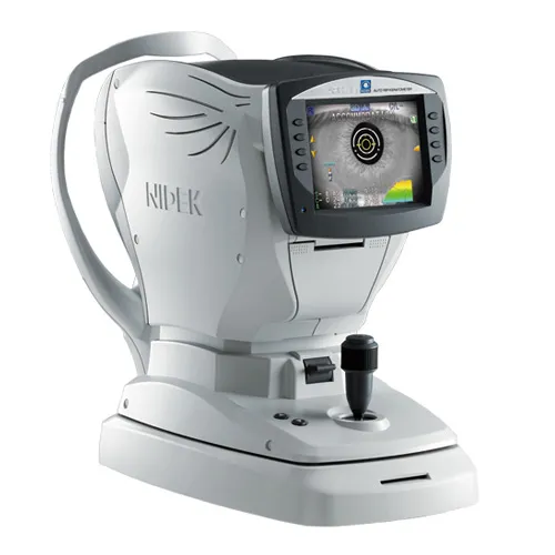
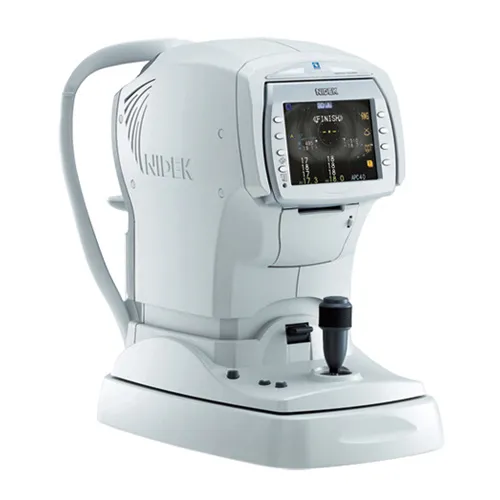
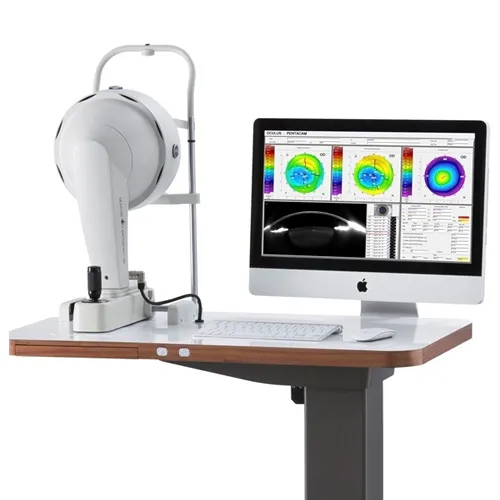
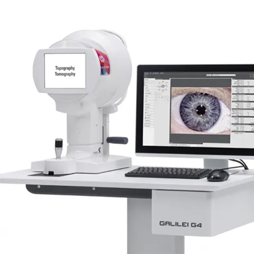
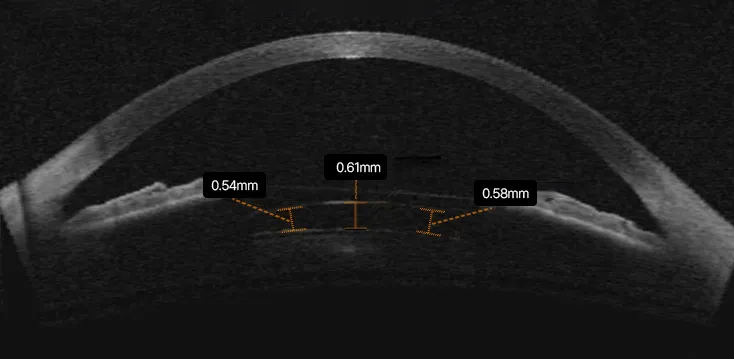
It covers detailed diagnostic examinations such as UBM, anterior segment OCT, vault prediction analysis, and internal eye measurements required to determine the correct lens size. It also includes customized lens preparation, surgical planning, and post-operative safety monitoring.

These steps are essential because accurate lens sizing directly affects long-term stability and helps reduce complications such as improper vault, cataract risk, or elevated eye pressure.

Choosing Surgery Based on Safety, Not Just Price

Choosing implantable collamer lens surgery based only on the lowest price may not always be the safest decision.

ICL surgery is not simply a procedure where a lens is inserted and finished. The most important part is the detailed planning before surgery — including precise measurements, lens selection, and safety evaluation tailored to the individual eye.

At EYE Plus Eye Clinic, treatment decisions are made based on long-term safety and eye health rather than price alone. Careful examination and proper surgical planning help minimize ICL surgery risk and support stable, lasting vision correction.

* More accurate cost guidance is provided after checking the individual's eye condition through detailed examination and consultation.

* If you are curious about what vision correction surgery suits your eyes, please feel free to consult or make an appointment through the button below.

Frequently Asked Questions About ICL Surgery

Consultation Request

Please leave your questions for EYEPLUS,
and our staff will contact you after reviewing them.